Each case of ringbone will be described as either high (pastern joint area) or low (coffin joint area), and by whether it involves the joint (articular) or not (periarticular). Traumatic ringbone may occur anywhere along the pastern. It’s important to make the distinction between the types of ringbone because this influences the prognosis for soundness with treatment.
Our ”Types of Ringbone” table (see above, right) lists the various types of ringbone and possible causes. Horses with club feet or upright pasterns are less efficient in distributing forces on impact and this jamming and jarring is hard on the pastern and coffin joints. Biomechanical studies have shown that the hoof wall and these two joints absorb most of the force of impact. Horses whose pastern and foot axis deviates in or out rather than being directly aligned under the fetlock may be at higher risk for ringbone. However, this risk can be minimized by trimming the hoof to keep the bones in their natural alignment.
More ringbone is caused by trimming that forces the foot back under the ankle, or deliberately introduces side-to-side imbalances to ”correct” winging in or winging out. ”Corrective” trimming can’t realign the horse’s natural bone axis. When trimming doesn’t follow the natural alignment of the bones, uneven forces are developed that can easily result in ringbone caused by joint, joint capsule, tendon or ligament damage.
When ringbone appears at an early age, the cause is often OCD (osteochondrosis). Defects in the formation of a healthy bone cartilage junction that occur while in the uterus or at a young age lead to joint disease and bone chipping that causes articular ringbone.
Risk Factors
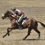
Sport: Jumpers, eventers, polo ponies and working Western horses, which must make sharp turns at high speeds, are at increased risk for ringbone both because of uneven joint loading and risk of soft tissue tears (joint capsule, tendons, ligaments).
Hoof Care:Poorly balanced hooves that cause the horse to land on one side of the hoof more than the other also are subject to uneven joint loading, with high forces through the joints on the side that lands first and excessive stretching of joint capsule, tendons and ligaments on the other side. Leaving the heels too high has also been shown to result in more movement of the pastern joint.
Concussion: Work on hard surfaces increases the forces on the lower leg joints on impact.
Diagnosis
Early ringbone, before much of anything is even visible on X-rays, can be an extremely painful stage for the horse. Conversely, seeing extensive calcium deposits on an X-ray doesn’t necessarily mean the area is causing the horse pain. As with any lameness, nerve blocks and/or local anesthesia of joints or areas of suspected soft-tissue damage must be done to confirm a diagnosis of lameness that is caused by ringbone.
Treatment Options
Available treatments depend on the location and type of ringbone, but the first step should be a search for correctable causes. Standing in front of the horse, check to see if the center of the toe corresponds to a line drawn directly through the middle of the bony column of the pastern. If it doesn’t, consult your farrier.
If you examine a hoof like this from the sole view, you’ll see that there is more sole on one side than the other, and the tip of the frog often does not point to the center of the hoof wall at the toe. On the side view, check for excessive heel height and any deviation from a smooth, straight axis through all the bones of the pastern and foot. X-rays can help with these evaluations, and a conference between your farrier and veterinarian to decide on needed corrections is in order.
Six to eight weeks rest, to allow active areas of inflammation and remodeling to quiet down, is usually recommended.
During this time, the horse may benefit from being barefoot if his hoof is stable and strong enough. Ideally, the frog will have contact with the ground when the horse is weightbearing to take full advantage of the natural shock absorbing structures of the foot. Toes should be short enough to allow easy breakover and rounded. If shoes are used, a beveled or rocker toe assists with breakover.
Many horses also benefit from either full pads with soft-support material underneath, Therapeutic Honeycomb pads from Supracor (www.supracor.com/, 888-924-6773), or the newer gel support pads from (www.impactgel.com/, 866-321-8729). The gel pads provide gentle, even support and mold themselves well to the contours of the individual foot.
Bottom Line
High periarticular ringbone, which involves areas of soft-tissue attachment to bone, has the best prognosis. Meticulous attention to trimming and hoof dynamics coupled with a sufficient amount of rest to allow the area to quiet down and set up may be all that is required to return the horse to soundness. If extensive damage and tearing to soft tissue structures stabilizing the joint has been diagnosed, several months rest may be needed to get good healing. Progress can be followed by ultrasound and X-rays. Some vets may even choose to put these horses in a cast for a few weeks, to further minimize movement. These horses may also respond well to shock-wave therapy.
Low periarticular ringbone is basically treated the same way, but it may not be accessible for shock-wave therapy. While the prognosis is better than for low ringbone that actually involves the coffin joint, it’s more difficult to protect this area from concussion.
Young horses with articular ringbone related to OCD fragments has a guarded prognosis. Arthroscopic surgery to flush the joint and remove the irritating fragments is usually necessary and may be followed by a prolonged course of Adequan, Legend, and/or oral joint nutraceuticals.
Articular ringbone always means underlying arthritis as well. It used to be thought that problems involving the pastern joint had a better prognosis because this joint was relatively immobile. However, newer studies have shown much more movement at the pastern joint than was previously thought.
Both high and low articular ringbone is usually initially treated as above (trimming, shoeing, rest) and with intra-articular injections of hyaluronic acid and/or corticosteroids. Many horses can continue to compete for many years with these treatments.
If it’s not enough to halt the process, surgery is an option. Screws and/or plates can be placed to bridge and stabilize the joint. Success rates as high as 85% have been reported. Unfortunately, there are no surgical options for low articular ringbone.