A University of Minnesota study reported that of 124 cases of suspected back pain seen at their clinic over five years, over half of the horses had a sacroiliac-area problem.
At the University of California, researchers examined the spines of 36 Thoroughbred racehorses euthanized for unrelated reasons and found acute injury or degenerative/arthritic changes in the sacroiliac joint in all the horses.
In the United Kingdom, the Animal Health Trust reported dressage and jumping horses to be high risk for sacroiliac region pain. Clearly, the sacroiliac region needs to be seriously considered any time a horse is off behind or has back pain.
What Is The Sacroiliac’
The sacrum is the end portion the horse’s spine. If you press a finger firmly along the middle of the horse’s back and move your finger back toward the tail, you’ll feel a prominent dip a short distance past the flank. This is the lumbosacral space and marks the spot where the lumbar vertebrae end and the sacrum starts.
The ilium is one of three bones that make up the pelvis. It’s shaped roughly like a wing, or the broad, flat section of a moose antler without the spikes, and is the most forward part of the pelvis.
The sacroiliac joint is the junction between the horse’s spine at the sacrum (“sacro”) and the pelvis at the ilium (“iliac”). The joint is buried under the heavy gluteal muscles of the top of the horse’s rump, lying just off the midline on both sides, on a line drawn between the back edge of the tuber sacrale and the spine. It sits at about the highest point of the rump. The tuber sacrale is the bony prominence just behind the flank that is commonly called the “point of the hip,” although it has nothing to do with the hip joint.
This area has two sets of ligaments, either of which can be damaged and cause pain. The dorsal sacroiliac ligaments run from the tuber sacrale (“point of the hip”) over to the top of the sacrum. They don’t involve the sacroiliac joint directly, but they do help anchor the ilium to the sacral spine.
The ventral sacroiliac ligaments are located deeper, in the area of the sacroiliac joint itself, which they stabilize. This assembly is designed to hold the horse’s pelvis tightly to his spine. A normal sacroiliac joint is capable of little movement and contains minimal amounts of joint fluid.
Causes Of Pain
Ligament injuries. Pain in the general area of the sacroiliac may involve injury to the ventral ligaments that stabilize the joint. In some cases, the injury may be an actual subluxation, although this isn’t as common as arthritis or ventral ligament strain/damage. The dorsal ligaments running from the tuber sacrale to the sacrum are also often injured, although this injury doesn’t necessarily also involve the sacroiliac joint itself.
Fractures. Stress fractures along the wing of the ilium have been found in racehorses, both microfractures and those severe enough to show up on X-rays. These fractures are presumed painful. Any of these, alone or in combination, can cause sacroiliac area pain.
Problems
By having a joint connecting his pelvis to the spine, rather than a solid bridge of bone, the horse gets the benefit of having tissues with a bit more flexibility to them, including some shock-absorbing capacity. However, the sacroiliac joint is mainly an anchor, not designed for much movement.
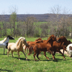
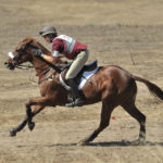
The joint can be overstressed by speed and/or extremes of movement of the hind leg. Racehorses are at risk, as are horses that work with a high degree of hind-end engagement, such as barrel racing, reining, jumping, dressage and horses that work over sharp inclines. Sacroiliac area injury may also occur if the hind leg suddenly slips — as could happen when on mud, ice, pavement and so on — and could even occur on turnout.
Diagnosis
There are no characteristic symptoms or signs that point to the sacroiliac area as a cause of lameness. A sacroiliac injury would be suspected on a horse with a hind-end lameness that can’t be localized to joints or soft tissues lower down the leg, such as when the horse had local anesthetic blocks up to and including the stifle without eliminating the lameness.
History may also play a role in making a diagnosis. You would suspect the sacroiliac in a horse that:
• Is involved in a high-risk sport/training program or was observed to slip and fall at pasture.
• Shows a loss of flexibility through the lower back.
• Can only manage a poor bascule over fences, jumps long, refuses jumps, stops in combinations.
• Has difficulty with cavaletti.
• Demonstrates a loss of impulsion on the flat, is difficult to collect, or “strung out” at the canter.
• Obviously prefers one lead and frequently cross-canters.
• Shows a reluctance or refusal to work tightly, perform sliding stops or turn on the haunches at speed.
In addition, you may note that the horse has:
• Hunter’s bump or knocked-down hip that is distinguished from an anatomical asymmetry or old injury no longer causing pain.
• Uneven muscling, a sore side with less muscle.
• Pain on palpation in the area of the tuber sacrale, which suggests either a fracture or involvement of the dorsal sacroiliac ligaments.
• An exaggerated sensitivity to palpation over the lower back and sacrum, which may be either a sinking away from the pressure, or tensing and quivering of the muscles (or a good swift cow kick).
• Pain at the extremes of range of motion when the leg is pulled back and/or forward. But this is nonspecific and only suggests a possible problem anywhere in the sacro-iliac area, pelvis, hip, even femur.
Diagnostic Options
Radiography: X-rays are of limited value both because of the difficulty in obtaining them (horse must be under anesthesia and on his back) and because there may be little to nothing to see in most sacroiliac area injuries. They are most indicated in horses suspected to have a fracture.Scintigraphy (“bone scan”): Although there can be some slight overlap in uptake patterns between horses with sacroiliac-area problems and normal ones, especially in older horses, bone scanning is an accurate way to confirm the diagnosis. However, it can’t pinpoint if the problem involves ligaments, joint or bone.
Thermography: Thermographic images of the back may reveal a “cold spot” at areas of ligamentous injury involving dorsal ligaments.Diagnostic Ultrasound: This can show thinning, thickening, scarring or hypoechoic areas in the dorsal ligaments. The joint can be viewed using an ultrasound probe in the rectum, which can pick up asymmetries from side to side.
Local Anesthesia: Your vet may be able to get in the neighborhood of the sacroiliac and the supporting ventral ligaments with a long needle. However, because the joint is so deep and shallow, it’s difficult to actually enter it and be sure anesthesia doesn’t go anywhere but into the joint. Therefore, local blocks can’t localize the problem to the joint itself, but a response does at least tell you that area is causing the pain.
Rectal Examination: Rectal examination can pick up asymm etry in the pelvis, localized swelling or thickening, sensitivity to touch, fractures. It’s probably an underused diagnostic technique.
Prognosis
Not surprisingly, the prognosis largely depends on the extent and nature of the problem. Because sacroiliac area injuries usually involve one or more ligaments, recovery will always be fairly prolonged, from six to eight weeks for problems that are caught early, up to many months.
Time is the first requirement for successful therapy. Ultrasound exams can provide detailed information on the extent of the injury, at least to the ligaments, and may help determine expected lay-up time. Serial exams can monitor healing progress.
First, you must get the inflammation under control. Your veterinarian may recommend a course of systemic anti-inflammatories (e.g. phenylbutazone, flunixin or herbal) or local injections (corticosteroid, herbal such as Sarapin, or homeopathic such as Traumeel). Because they can slow healing and result in ligament weakness, corticosteroid injections shouldn’t be done repeatedly, but their judicious use with severe inflammation may be warranted.
After the initial inflammation is under control, the horse should be kept moving. (Note: Complete stall confinement with restriction of movement is rarely necessary, except with fractures or a rupture of the ligament.) Field turnout is best, as long as the footing is good, with no steep inclines to negotiate and no other horses in the group that could force the injured horse to exercise more than he would voluntarily.
If field turnout isn’t an option, the horse should at least have as large a stall/pen as possible, liberal paddock time and be hand-walked. Some veterinarians also recommend daily range of motion exercises, moving the leg as far forward and as far back as comfort allows.
Long-term use of pain medications or corticosteroids should be avoided, so that progress can be monitored accurately and the horse doesn’t exercise more than he should.
Some veterinarians will use periodic Traumeel or Sarapin injections to help with low-grade pain/inflammation during rehab, as it may help the horse work through the rehab program to achieve the best possible flexibility. Injection of counterirritants such as iodine used to be popular, but it’s probably best avoided because it can increase scarring in the area.
Bottom Line
The truth of the matter is that most horses with sacroiliac-area injuries, even fractures, can return to full use if given enough time to heal. Ligaments heal the slowest of all tissues, and nothing can change that.
The oldest effective treatment is turnout for nine months to a year. When you see the horse is sound again at all gaits at pasture, it’s time to gradually resume formal work to bring him back.
Although severe injuries certainly could limit a horse’s future career, especially in demanding sports, many horses that reportedly fail to return to their original work do so simply because they aren’t given enough time.
Also With This Article
“Put It To Use”
“Minimize The Chance Of Injury”
“The Scoop On Alternatives”
“Hunter’s Bump”